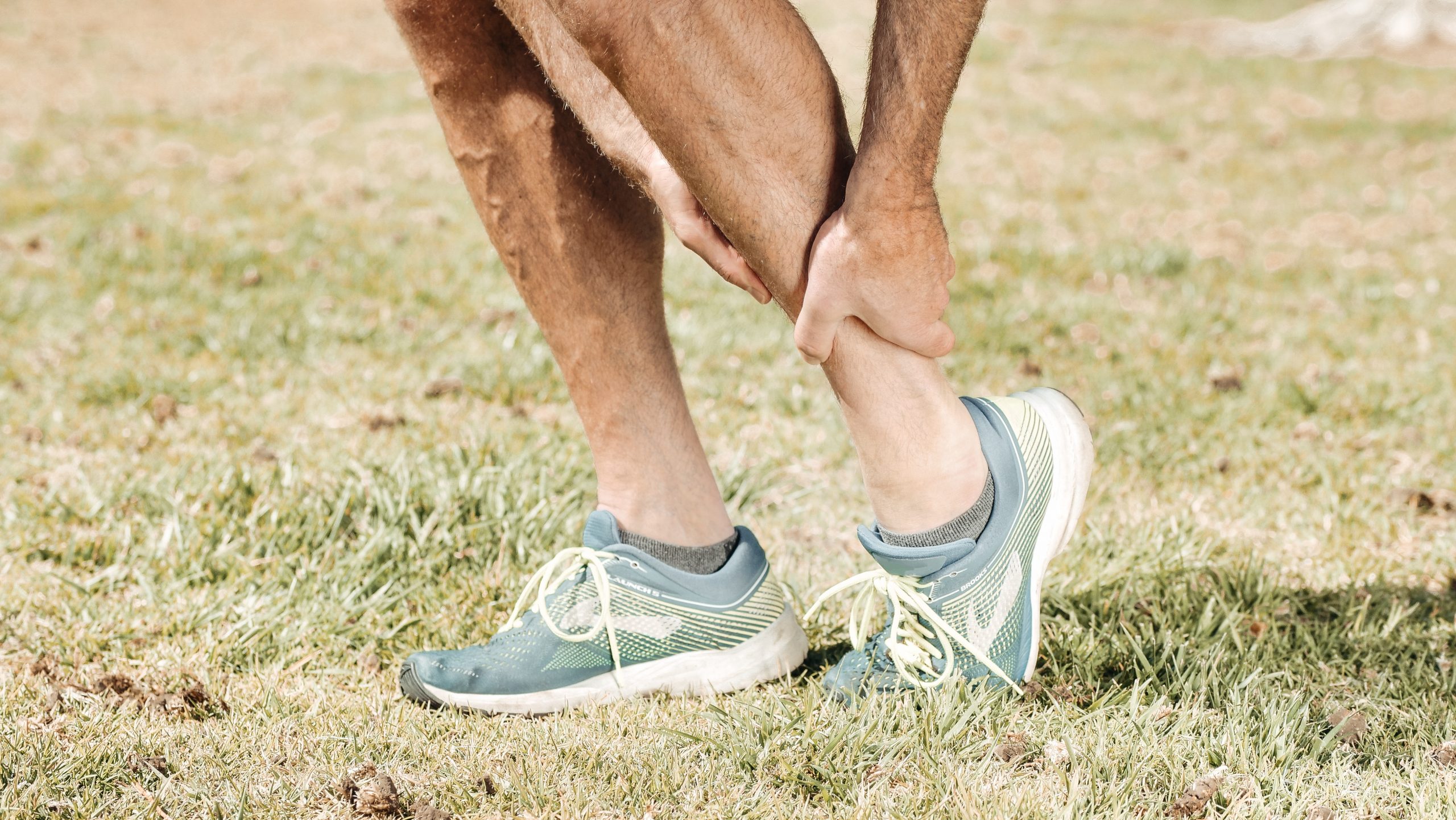
Fractures from fatigue in sports are common injuries in athletes and can have a major impact on the rhythm of training and competitions. The lower extremity is one of the most frequently affected, with overuse damage to the bone due to repeated and one-sided stress.
In general, a distinction is made between two types of fatigue fractures, those in which the healthy bone is overloaded and those in which a fatigue fracture occurs under normal stress but with a weakened bone structure.
The greatest influence on the development of fatigue fractures is caused by training stress and strain. Symptoms are mostly unspecific, typically appear during exercise and can lead to the discontinuation of physical activity. Soft tissue swelling or callus formation can occur in the late stages.
Radiologically, changes are only visible late. The MRT examination has established itself as the gold standard for early detection and staging appropriate to the stage. The type and duration of treatment and rehabilitation can be determined accordingly.
The central pillar of the treatment of fatigue fractures is the reduction of the (training) strain to a level below the pain threshold. In principle, surgical therapy is rarely indicated, but there are specific fatigue fractures in which conservative management can be frustrating.
Background
Fatigue fractures are common injuries among athletes and occur predominantly in the feet and legs. The tibia, navicular bone, metatarsal bone, and fibula are predisposed.1 This mostly affects runners or athletes, but games such as soccer, volleyball or basketball can also provoke fatigue fractures. Fatigue fractures are also observed in the upper extremities, e.g. stress fractures of the carpal bones in tennis or squash players. The frequency of fatigue fractures is very variable depending on the sport and can reach up to 47%. 2, 3
Fatigue fractures (= stress fractures) are damage to the bone due to repeated, one-sided stress, which overtaxes the bone due to its uniformity and durability. This leads to changes in the arrangement of trabeculae (which represent the supporting lines of the bone) and subsequently to a reaction of the periosteum in the form of fluid accumulation (edema). The accumulation of fluid (bone marrow edema) is usually the point in time at which pain occurs for the first time.
A basic distinction is made between two types of fatigue fractures, those in which the healthy bone is overloaded and those in which a fatigue fracture occurs under normal stress but with a weakened bone structure. The greatest influence on the occurrence of fatigue fractures is caused by training stress and strain, which is why the analysis of stress, training conditions and risk factors is necessary.
Clinic
The symptoms of fatigue fractures are mostly non-specific. The main symptom is pain which is often described as dull and typically occurs during exercise. In the initial stage, exertional pain only occurs towards the end of the sporting activity, in the advanced stage the onset of pain shifts steadily to an earlier point in time and can ultimately even lead to the discontinuation of the sporting activity. Recent changes in training (increase in intensity, duration) or extrinsic factors (change of footwear or surface) should be inquired about based on anamnesis.
In the late stages, soft tissue swelling or callus formation can be seen on clinical examination. Provocation tests (with compression / flexion) can provide evidence of the presence of a fatigue fracture. The mobility of the adjacent joints is usually not restricted and painless. The search for anatomical misalignments that can favor a stress fracture (e.g. genu varum, flat or hollow feet, leg length differences) must be carried out.
Imaging
Imaging methods are required for a reliable diagnosis and staging appropriate to the stage.
In conventional X‑rays, changes in the bone structure in fatigue fractures become visible late or not at all. Typical signs here are the elevation of the periosteum, thickening of the cortex, callus formation or, finally, the marking of a fracture line.4 If the x‑ray image is normal but the clinical suspicion of a fatigue fracture, the diagnosis with magnetic resonance imaging (MRI) or bone scintigraphy should be escalated.5–7 Several studies have shown that MRI is equivalent to bone scintigraphy in terms of sensitivity, but has an even higher specificity. 5–7
The MRI examination is used to classify the severity according to Arendt et al.6, which classify grades 1 and 2 as “low grade” and grades 3 and 4 as “high grade”. Fredericson et al.7 also differentiate in their classification between periosteal and bone marrow reactions. This staging shows a good correlation between radiological findings and clinical symptoms in everyday clinical practice. Based on this classification, further procedures and duration of treatment can be determined. Another advantage of MRI is the low radiation exposure, which enables follow-up examinations at intervals.
Possible differential diagnoses for a fatigue fracture — in addition to osteomyelitis and osteon corps — are bone contusion with a so-called “bone bruise”. The differentiation in the MRI is often difficult, pointing the way here are trauma history (direct trauma during contusion) and occurrence near the joint.
Therapy
A detailed anamnesis is crucial for the success of the therapy when diagnosing a fatigue fracture. Inquiring about and identifying possible training errors, changes in training intensity, failure to take account of changes in extrinsic factors (surface, footwear), lack of rest breaks or other injuries are key factors here.
The main pillar of therapy is reducing physical activity to a level below the painful level and modifying training. In the clinical course, pain has proven to be the best indicator for the start of therapy / the healing process. The doctor-patient relationship is another key point. Detailed information about the clinical picture and course and the necessary compliance of the athlete are necessary in order to avoid the risk of the fatigue fracture progressing to the next stage or to a complete fracture.
Conservative therapy comprises three phases as summarized by Albrecht et al.1:
- Pain control by means of cooling, physiotherapeutic measures, training breaks or modified training breaks.
The (partial) relief on forearm crutches is necessary if pain is already present when walking in everyday life. The later increase in load should be decided on the basis of the pain progression. If there is no pain, the load can be increased in a controlled manner, with a day of rest for regeneration after exercise. Phase (2) begins after 3–5 painless days.
- Exercises with loads, but without impacts (e.g. steppers) and sport-specific muscle training. Muscular imbalances should be corrected during this phase. Endurance training such as swimming, aqua jogging or cycling can be done if it can be carried out painlessly.
- The last phase provides for the dosed, continuous return to sport-specific activities.
Basically, most fatigue fractures respond to a conservative therapy regimen; surgical therapy is rarely indicated. However, there are fatigue fractures that show an increased risk of delayed bone healing, development of pseudarthroses or the formation of a complete fracture. An example of this is the anterior tibial stress fracture. The surgical therapy options available are intramedullary nailing, drilling of the pseudarthrosis and debridement with spongiosaplasty. 3, 8, 9
Conclusion
Fatigue fractures are common injuries among athletes. In the case of diffuse stress pain in the area of the bone during sporting activities, a stress fracture should be considered, all the more if there has recently been a history of a change in training habits. If the diagnosis is confirmed at an early stage, a few weeks of rehabilitation time are usually sufficient to return to familiar training and competition conditions. In the event of a clinical suspicion of a fatigue fracture, the diagnosis should therefore be escalated with an MRI (= gold standard) at an early stage. Most fatigue fractures respond to conservative therapy management, but surgical therapy is rarely indicated.
Authors: Univ.-Prof. Dr. med. Ulrich Stöckle and Priv.-Doz. Dr. med. Lucca Lacheta
Correspondence address:

Univ.-Prof. Dr. med. Ulrich Stoeckle
Charité — University Medicine Berlin
Augustenburgerplatz 1, 13353 Berlin
Email: ulrich.stoeckle@charite.de
Literature
1. Albrecht SB, R.M. Stressfrakturen. Switzerland. Zeitschrift für Sportmedizin und Sporttraumatologie. 2004;52:27–30.
2. Bennell KL, Malcolm SA, Thomas SA, Wark JD, Brukner PD. The incidence and distribution of stress fractures in competitive track and field athletes. A twelve-month prospective study. Am J Sports Med. 1996;24:211–217.
3. Chang PS, Harris RM. Intramedullary nailing for chronic tibial stress fractures. A review of five cases. Am J Sports Med. 1996;24:688–692.
4. Harrast MA, Colonno D. Stress fractures in runners. Clin Sports Med. 2010;29:399–416.
5. Deutsch AL, Coel MN, Mink JH. Imaging of stress injuries to bone. Radiography, scintigraphy, and MR imaging. Clin Sports Med. 1997;16:275–290.
6. Arendt EAG, H.J. The use of MR imaging in the assessment and clinical management of stress reactions of bone in high-performance athletes. Clin. Sports Med. . 1997;16:191–306.
7. Fredericson M, Bergman AG, Hoffman KL, Dillingham MS. Tibial stress reaction in runners. Correlation of clinical symptoms and scintigraphy with a new magnetic resonance imaging grading system. Am J Sports Med. 1995;23:472–481.
8. Green NE, Rogers RA, Lipscomb AB. Nonunions of stress fractures of the tibia. Am J Sports Med. 1985;13:171–176.
9. Orava S, Karpakka J, Hulkko A, et al. Diagnosis and treatment of stress fractures located at the mid-tibial shaft in athletes. Int J Sports Med. 1991;12:419–422.
